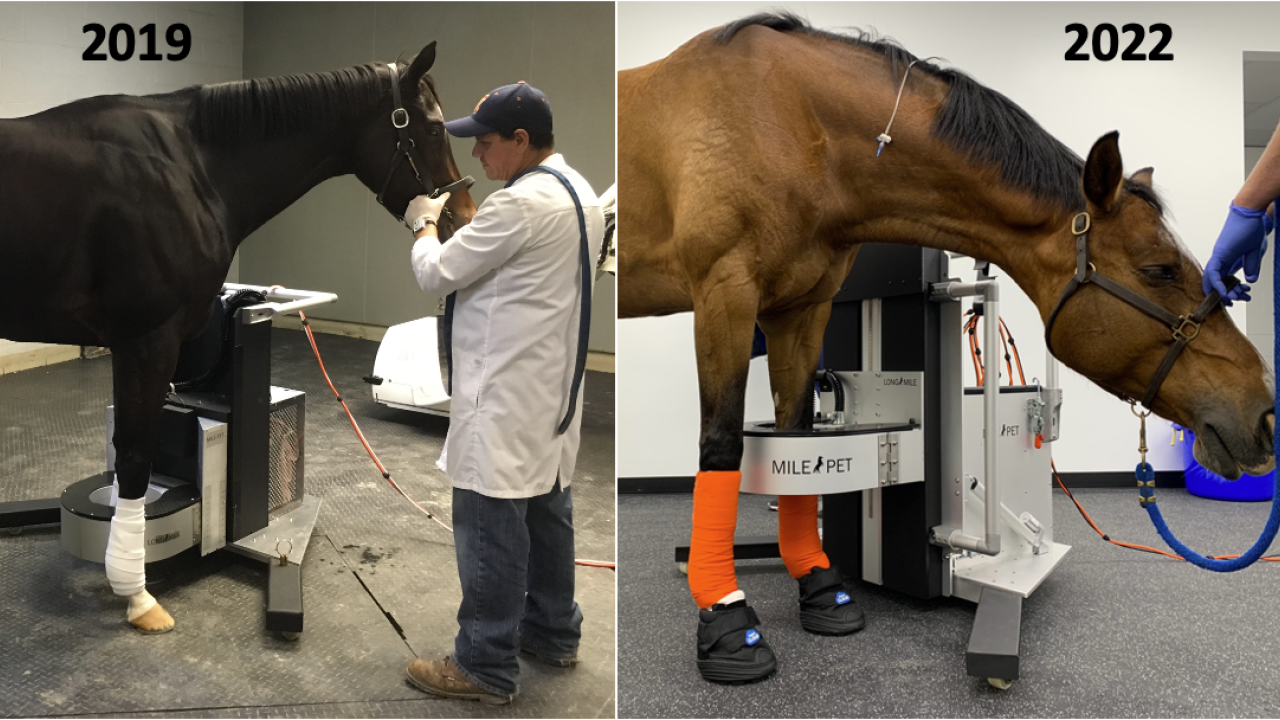
Standing Equine PET Going Strong as a 3-Year-Old
Now in its third year of application at Santa Anita Park, positron emission tomography (PET) scans have benefited more than 500 horses at the renowned racing facility.
This novel imaging modality, pioneered at the UC Davis School of Veterinary Medicine, was initially focused on the racehorse fetlock, but has matured in the last year. The early equine PET studies performed on racehorses at Santa Anita have now been published and have confirmed the value of this new technique. PET was found superior to bone scan in assessing the racehorse fetlock, particularly for identifying injuries of the sesamoid bones, the most common cause for catastrophic breakdowns at the racetrack. PET also demonstrated its ability to monitor injuries over time and predict the amount of time needed to heal.
A multicenter study, combining in addition to Santa Anita horses, racehorses from Golden Gate Fields (imaged with the UC Davis PET scanner) and racehorses from the Fair Hill Training Center (imaged with the PennVet New Bolton Center PET scanner), has looked into what can be found with PET in horses racing successfully. This study represented an important steppingstone in the development of screening strategies to identify horses at risk for catastrophic injuries. A summary of this work was presented at the Welfare and Safety of the Racehorse Summit organized by the Grayson-Jockey Club Research Foundation.
The availability of the technology has significantly increased in the past year. In the first two years of standing equine PET, only three scanners were available (Santa Anita, UC Davis, and PennVet). Four more scanners were installed in 2022 with Rood and Riddle Equine Hospital adding the scanners at their Lexington (KY) and Wellington (FL) locations. Additionally, two other scanners are now operating in Florida at the World Equestrian Center Hospital, operated by University of Florida in Ocala, and the Ocala Equine Hospital. Three more installations are planned in the United States, including Churchill Downs, resulting in a total of 10 different sites equipped with the technology by the end of 2023.
With this increased availability comes the access to more diverse populations of horses and a broadening of applications of the technique. Beyond racehorses, many pleasure and sport horses also now benefit from the use of PET. Standing PET can be used from the foot to the knee in the front limbs and from the foot to the hock in the hind limbs.
Dr. Mathieu Spriet, professor of diagnostic imaging at UC Davis, recently presented a summary of the various applications at the 2022 American Association of Equine Practitioners Convention.
“Standing equine PET has revolutionized our approach to imaging of lameness,” summarized Spriet. “PET can be used as an advanced imaging technique in addition to CT or MRI in complex cases, but in other cases it can simply be combined with x-rays and ultrasound providing an affordable alternative to other advanced imaging.”
Spriet’s work shows common applications of PET for bone injuries, in particular of the foot, fetlock, and hock, but also for soft tissue injuries in tendon and ligaments. On-going studies are investigating the values of PET on other important topics such as laminitis and infection.
# # #
