
Surgery Saves Puppy in Congestive Heart Failure
“Case of the Month” – October 2021
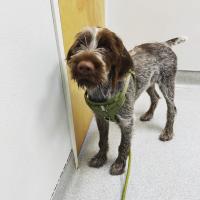
When George William, a now 6-month-old wirehaired pointing Griffon, was brought to his first veterinary appointment as a new puppy, a heart murmur was discovered. His general practitioner told owners Teresa and Paul Struffert that it was very serious, and they needed to take George William to the Cardiology Service at the UC Davis veterinary hospital.
Cardiology residents Drs. Ashley Walker and Ashley Sharpe worked with faculty members Drs. Joanna Kaplan and Marisa Ames to diagnose George William with a patent ductus arteriosus (PDA), a birth defect that causes heart failure.
A PDA results in the persistence of the ductus arteriosus, a vascular structure important during development in utero. The ductus arteriosus is a small channel that connects the pulmonary artery (which carries blood to the lungs after birth) and the aorta (which carries blood to the rest of the body). In the womb, it is responsible for conveying blood past the non-functional lungs (since puppies don't breathe air before birth), and into the systemic circulation. At birth, when an animal takes its first breath, the lungs become filled with air. This causes a decreased resistance to blood flow, and blood moves through the blood vessels of the lungs, instead of through the ductus arteriosus. In the normal animal, the ductus arteriosus should fully close after birth. When the ductus arteriosus does not close, extra blood continuously circulates through the lungs and the left side of the heart which can result in heart failure (fluid in the lungs) and death by one year of age.
To close George William’s ductus arteriosus, the cardiologists had to stop blood flow through the PDA. There are two ways to achieve this – either through an open-chest surgery to manually tie off the ductus, or by a minimally invasive interventional procedure using a catheter to place a small device inside the ductus to stop blood flow. In the latter method, cardiologists feed a long catheter through the femoral artery and into the aorta (main artery in the body). Once corrected, prognosis for survival is good, with many patients living a normal lifespan.
In George William’s surgery—assisted and monitored closely by the Anesthesia/Critical Patient Care Service—the team utilized the minimally invasive implantation of an Amplatz Canine Ductal Occluder to occlude the PDA. George William recovered wonderfully from the surgery and was discharged the following day.
Teresa reported it was not easy to keep him inactive for several weeks, as it would be for any young puppy, but her family closely followed his recovery instructions. At his recheck appointment, George William was doing great, and his congestive heart failure had resolved.
Since recovering fully, George William has been doing a great job at learning commands, and Teresa is interested in making him a therapy dog due to his friendly personality.
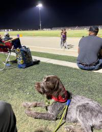
George William now enjoys spending his fall Friday nights at the local high school football games, where the Strufferts’ daughter is a cheerleader.
While more popular in parts of Europe where they were originally bred to be hunting dogs, the wirehaired pointing Griffon is still relatively rare in the United States.
# # #
George William's case showcases an example of cutting-edge and life-saving procedures that will be a hallmark of the future Veterinary Medical Center at UC Davis. As the final phase of the VMC, an entirely new Small Animal Hospital will be constructed, expanding the size and scope of the current facility. This will allow clinicians to expand their innovative procedures and continue to push the limits of veterinary medicine.