UC Davis Welcomes Equine Surgeon and Orthopedic Researcher Dr. Heidi Reesink
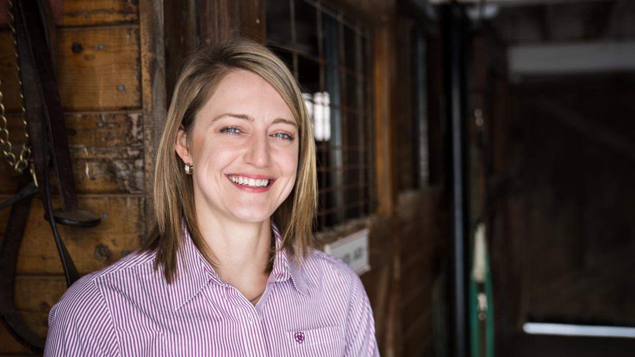
The UC Davis School of Veterinary Medicine is pleased to welcome Dr. Heidi Reesink as a Professor of Veterinary Orthopedics. A board-certified surgeon, Dr. Reesink will have a clinical appointment with the Veterinary Medical Teaching Hospital’s (VMTH) Equine Surgery and Lameness Service, as well as a research laboratory within the school’s J.D. Wheat Veterinary Orthopedic Laboratory.
Dr. Reesink received her VMD (Summa Cum Laude) from the University of Pennsylvania in 2007. She stayed at Penn Vet’s New Bolton Center to complete a 1-year internship in 2008 before completing a 3-year residency in large animal surgery at Cornell University in 2011. Dr. Reesink is a Diplomate of the American College of Veterinary Surgeons (Large Animal). Additionally, she received a PhD in Comparative Biomedical Sciences from Cornell in 2016.
She served as an Assistant Professor at Cornell from 2016-2022 and an Associate Professor from 2022-2025 before starting at UC Davis in March 2025.
Dr. Reesink’s research focuses on the pathophysiology and treatment of musculoskeletal disease, including osteoarthritis, tendinopathy, and fracture. Her laboratory seeks to advance One Health therapeutic design and interventional strategies aimed at restoring cartilage health, improving comfort, and prolonging joint longevity and patient mobility for both veterinary and human patients alike.
“One thing that drew me to UC Davis in particular is the collaborative nature of the university, both clinically and in the laboratory,” said Dr. Reesink. “We have the opportunity to study naturally occurring diseases in animal patients as models for the same diseases in humans, with tremendous potential benefits for both species.
“I’ve already met with researchers at the medical school to discuss how we can work together in researching similarities between horses, dogs, and humans. Collectively, we can learn more about the commonalities of joint disease in terms of identifying biomarkers, monitoring disease progression, and guiding targeted treatments.”
An example of this is soon-to-be-published research by Dr. Reesink on a comparative study of synovial fluid in anterior cruciate ligament (ACL) injuries in humans and animal patients.
“We found that the protein most upregulated in joint fluid in ACL injuries was the same in dogs and humans, and by a large margin,” said Dr. Reesink. “We’re excited to study that protein (periostin) further to evaluate whether it is promoting fibrosis in ACL injury and if it might be a therapeutic target for new joint disease medications.”
Additionally, Dr. Reesink is researching in vitro cell culture methods to study treatments for arthritis and understand the pathways that are altered in the disease and how the treatments might target those. She is also partnering with biomedical engineers to develop potential joint disease therapeutics specific to horses and dogs.
Clinically, Dr. Reesink emphasizes the importance of imaging and the excellence that UC Davis has achieved in that field, specifically utilizing positron emission tomography (PET scan) and computed tomography (CT scan) to maximize outcomes for equine patients.
“So much of what we are able to accomplish as clinicians is interconnected with imaging,” said Dr. Reesink. “We can make better decisions about a horse’s outlook if we can continually monitor its condition over time (with imaging) to see if the treatment or surgery was effective or to see if a fracture is healing properly.”
UC Davis will soon open the All Species Imaging Center at the VMTH. This central hub of imaging will include the hospital’s first standing equine CT scanner, which will allow veterinarians to image horses using only mild sedation rather than full anesthesia needed for a traditional CT scan.
“We are much more likely to achieve that continual monitoring by sedating a horse and walking it into a standing CT,” stated Dr. Reesink explaining how important it is to perform follow-up imaging to chart progression of recovery.
She also commented on the expanded capabilities of a standing CT scanner to capture more of the head and neck of a horse than most traditional CT scanners, in addition to the back and pelvis in an anesthetized horse. The All Species Imaging Center is slated to open this summer at the VMTH.
# # #