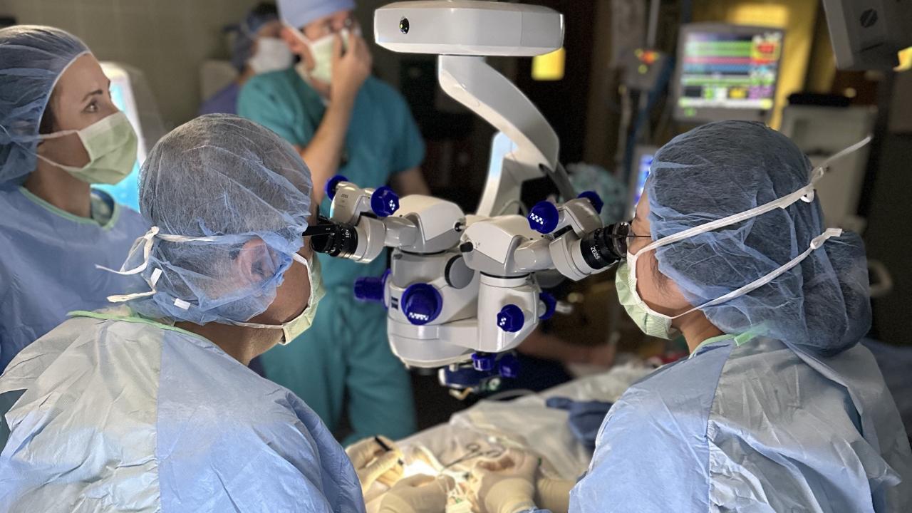
Equipment Upgrade Increases Ophthalmology Offerings
Cataract Surgery Cost Now Reduced by an Average of 20%
The Ophthalmology Service recently upgraded its surgical microscope, allowing the opportunity for a never-before-performed surgical procedure at the UC Davis veterinary hospital. This cutting-edge ophthalmic technology also opens more appointment opportunities, increased specialty training opportunities for residents, and an advanced approach to compassionate care.
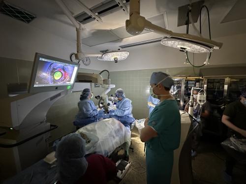
The technology of the new microscope is far more advanced than the service’s previous microscope. There are many upgrades with regard to the functions within it. For example, the new microscope has an advanced imaging component—optical coherence tomography (OCT)—that allows for near-histologic detail images (microscopic images used to help diagnose disease) to be taken of the eye while performing surgery. Previously, UC Davis veterinary ophthalmologists relied on OCT only as a pre-surgery assistance tool. OCT can pinpoint where exactly a foreign body is located in the cornea, and it can visualize specific areas of the retina and optic nerve.
OCT is a newer technology that is not available in many veterinary ophthalmology practices. The technology has just recently become available for intraoperative purposes. For teaching hospitals like UC Davis, the advantages of advanced equipment with upgrades like OCT are boundless for students and residents.
“OCT not only helps with patient care, but the teaching aspect of it is tremendous,” said Dr. Kathryn Good, chief of the Ophthalmology Service. “When you’re guiding residents with instructions like, ‘make an incision 5% deep within the cornea,’ you can actually see on the screen how deep a lesion might be or how deep your incision is thanks to the OCT technology. From a resident mentoring perspective, it’s a huge addition to our microsurgical training program.”
The microscope is dual headed, so two people can look through the scope at the same time, as an added teaching component benefit. Faculty veterinarians can see a resident’s work closely, and vice versa. The entire microscope unit comes equipped with a large screen to project what is being seen through the microscope so residents and students not directly participating in the surgery can still observe the procedure.
Areas of focus for new surgical technology:
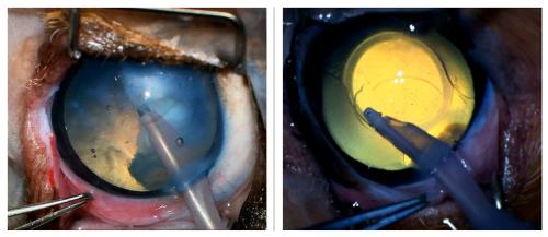
Right: Intraoperative image of cataract surgery demonstrating successful lens extraction and artificial intraocular lens implantation (note the artificial lens in place to restore visual acuity)
Cataract Surgery
Our ophthalmology specialists have had tremendous success with cataract surgeries for many years, and the new microscope is taking that success to the next level. Surgical dynamics and depth perception are better understood with the new equipment’s three-dimensional visualization. These advancements have streamlined our operating room workflow and increased appointment opportunities for cataract surgeries. Due to this, we have been able to lower the cost of cataract surgeries by up to 20% (costs may vary based on individual case needs).
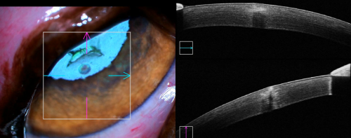
Foreign Body Removal
Whether corneal or intraocular, foreign body removals can now be performed more efficiently thanks to the microscope’s ability to utilize OCT during surgery, rather than previously only being able to rely on pre-surgery OCT. By integrating OCT into the surgical procedure, a real-time dimension for viewing structures of the eye immediately assists with decision-making during surgery. OCT images are optionally displayed in double-depth resolution in the surgeon’s view to better visualize small details like the location of a foreign object relative to a vital part of the eye.
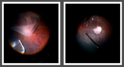
Right: Intraoperative image of laser retinopexy demonstrating successful retinal reattachment and visualization of the optic disc (note white dots indicating laser treatment sites)
Retinal Reattachment Surgery
An additional feature of the microscope unit is a back-of-the-eye viewing system that offers surgeons a clear and uncompromised view of the retina and other anatomical areas of the rear of the eye. This makes retinal reattachment surgery available now for the first time at UC Davis. Previously, clients had to travel to Southern California (or to only a handful of other clinics nationwide) for the surgery to regain vision lost by the detachment. The high-resolution visualization of the far periphery of the eye accentuates anatomical details, extends the depth of the field, and helps surgeons observe surgical maneuvers in the retinal tissue. Now, with the technology to perform this procedure, UC Davis is the only veterinary ophthalmology residency that has retinal reattachment as part of its training program. Previous training for veterinarians had to come from human medicine ophthalmologists. In fact, an ophthalmologist from UC Davis Health joined the service for the first procedure on a dog.
In addition to these three procedures, the microscope has proven valuable in other cases: corneal transplants, sequestrum in cats, the removal of necrotic corneal tissue, corneal tumors, and infected ulcers. The Ophthalmology Service also looks forward to using the microscope in future clinical trials of new procedures that work toward improving the vision of animals.
# # #
